MEGA-IOM

Multimodal System for Intraoperative Neurophysiologic Monitoring.
Unmatched integrated control of central and peripheral nervous systems to reduce postoperative risk and to improve surgical outcomes.
Rugged, durable, and exceptional usability.

Mega-IOM

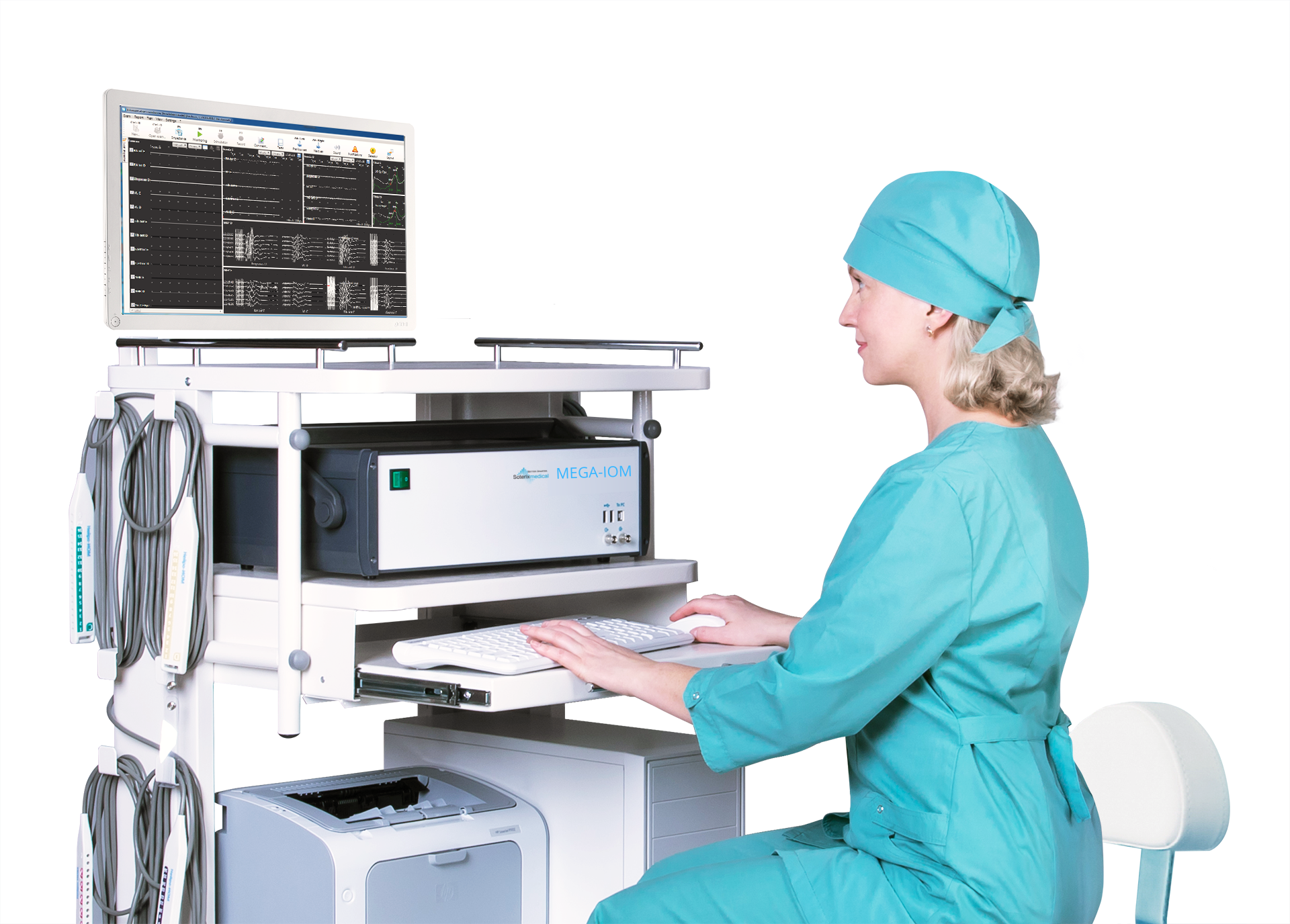
MEGA-IOM is a cutting edge modular platform solution that combines years of knowledge and experience obtained in clinical and research environments. Due to flexible configuration options the system can meet the most exacting needs of both surgeon and advanced neuromonitoring specialist.

The high performance system ensures full flexibility to the user. The predefined configurations are intended for almost all types of surgery procedures and ensure a simple workflow thanks to an intuitive user interface. Any parameter can be changed on-the-fly during recording and can be easily interpreted. The time-synchronized video and audio data contribute to obtaining reliable and accurate results.

A Comprehensive Solution for the Operating Room

The MEGA-IOM system ensures full featured performance due to the high quality and low noise signal acquisition, configurable display options, and auditory and visual feedback. The modern design with color-coded amplifier outputs and pods allows a specialist to speed up the startup procedure.

System Architecture

The MEGA-IOM system consists of an electronic unit with built-in amplifiers and stimulators, several pods with 5-meter extension cables, a PC, set of special electrodes, and dedicated software.

IOM Modalities

The MEGA-IOM system is delivered with preset templates ensuring the following modalities: free-run EMG, direct nerve stimulation including pedicle screw test, SSEP, MEP, EEG, AEP, VEP, ECoG, direct cortical stimulation, and train-of-four (TOF) stimulation.

Options:
Anesthesia depth monitoring, SpO2 recording

Practical Application of Modalities:

  • Motor evoked potentials (MEP) to evaluate the integrity of motor pathways.
  • Somatosensory evoked potentials (SSEP) to evaluate the integrity of sensory pathways and detect brain and spinal cord ischemia.
  • EEG to record and analyze brain activity (functional state of brain).
  • Direct stimulation to control the functional integrity of peripheral nerves.

Multimodal Intraoperative Neurophysiologic Monitoring

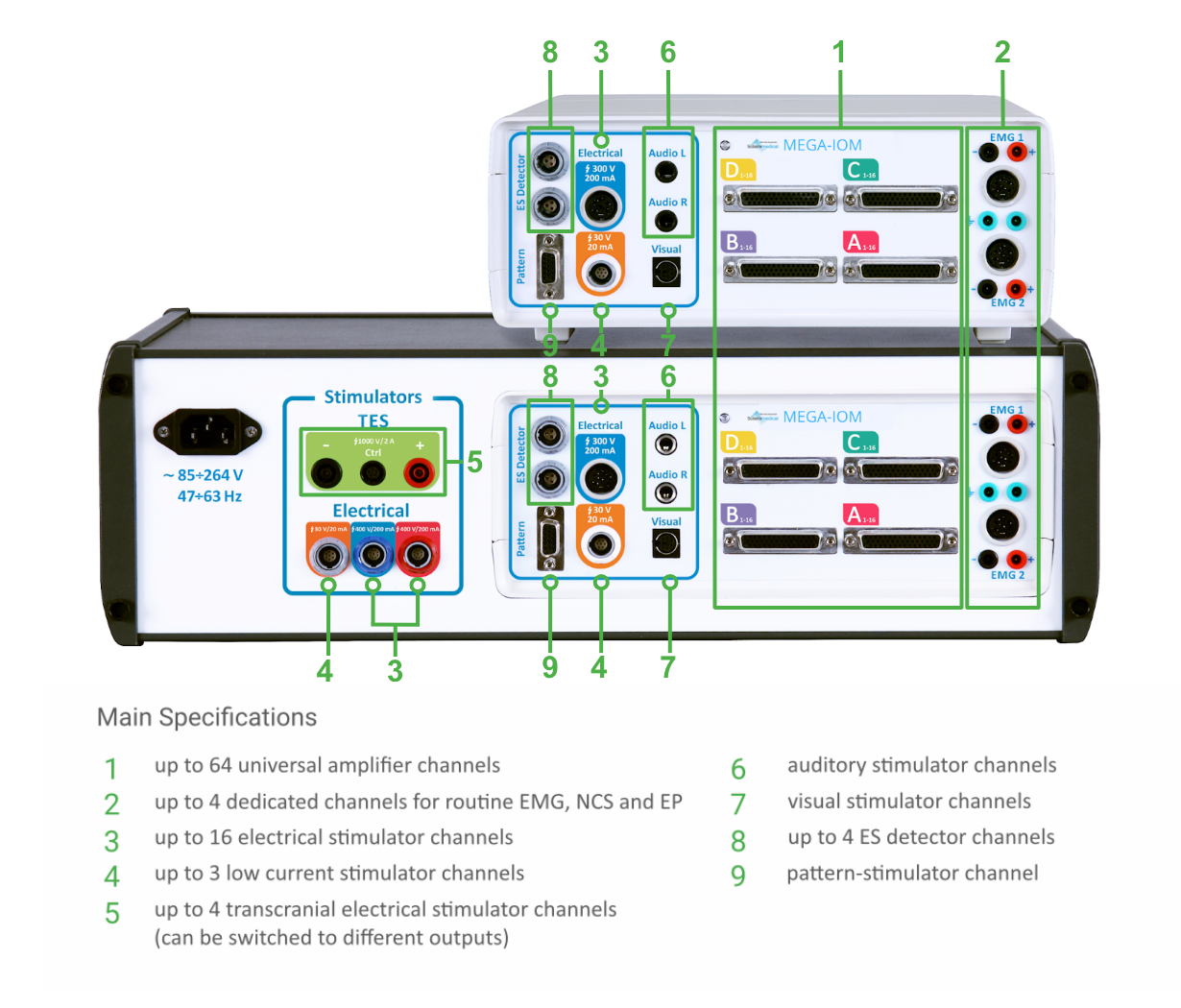
Transcranial Electrical Stimulator

For stimulation of the motor cortex in order to elicit motor evoked potentials (MEPs) in peripheral muscles. MEPs allow a specialist to monitor the integrity of motor pathways.

An electronic switch with 4 pairs of outputs to attach the stimulation electrodes is supplied. A pair of outputs to deliver a stimulus can be chosen in the MEGA-IOM.NET software.

Low Current Stimulator

For direct cortex stimulation, direct nerve stimulation, and pedicle screw test. Direct nerve stimulation allows the specialist to get direct feedback concerning nerve location. Direct cortex stimulation allows mapping of the eloquent brain areas.

A pod for attachment of different electrodes for nerve stimulation is supplied. A wide range of probes can be used: bipolar, monopolar, concentric, hook, etc.

8-Channel Electrical Stimulator

Allows for stimulation of up to 8 peripheral nerves simultaneously or in any sequence to elicit somatosensory evoked potentials (SSEP). SSEPs are sensitive to peripheral nerve events, spinal cord pathway injuries, and regional blood flow changes.

The system is equipped with 2 electrical stimulator pods (for channels 1-4 and 5-8). Each pod has 4 pairs of outputs that can function simultaneously.

Amplifier

The system is equipped with a reliable low-noise universal neurophysiological amplifier allowing high quality acquisition of EMG, EEG and multimodal EPs. The system can be supplied with 16-, 32-, or 64- bipolar channel amplifiers upon request.

The system is equipped with several pods with 5-meter shielded cables to attach the recording electrodes.

IOM Applications

Spine Surgery

  • Cervical/thoracic/lumbosacral decompression surgery
  • Pedicle screw positioning
  • Intra-/extramedullary spinal tumors
  • Scoliosis surgery
  • Tethered spinal cord syndrome
  • Dorsal rhizotomy

Vascular Surgery

  • Aorta Surgery
  • Blood vessel replacement
  • Endarterectomy
  • Heart Surgery

Brain Surgery

Intraoperative brain and cranial nerve monitoring. Mapping of functional areas of brain cortex and brainstem:

  • Brain tumors (and other abnormalities in motor, sensory, auditory, and visual cortical areas and their pathways)
  • Facial nerve monitoring
  • Epilepsy surgery
  • Recurrent laryngeal nerve monitoring during neck and thyroid surgery
  • Movement disorder surgery
  • Chiasmo-seller area (CSAB) monitoring
  • Aneurysm clipping

MEGA-IOM.NET Software

Monitoring Start

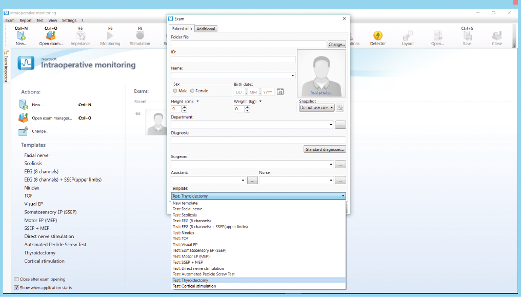
To begin monitoring, enter patient’s data and choose one of the test templates. The system provides a set of default templates for various types of surgeries.

Start Monitoring Image

Test Template Editor

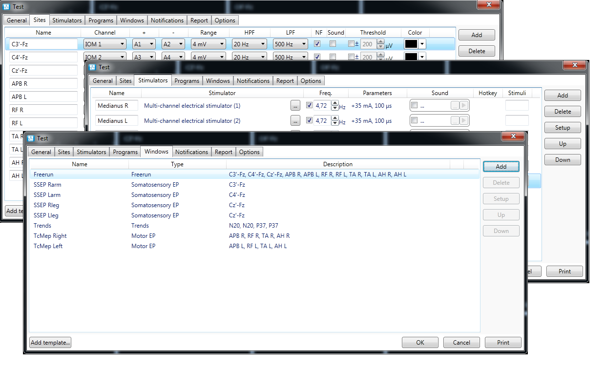
You can create your own test templates or edit existing ones by selecting stimulation and recording sites and adjusting the stimulation parameters for the current clinical case.

Test Template Editor image

Data Windows

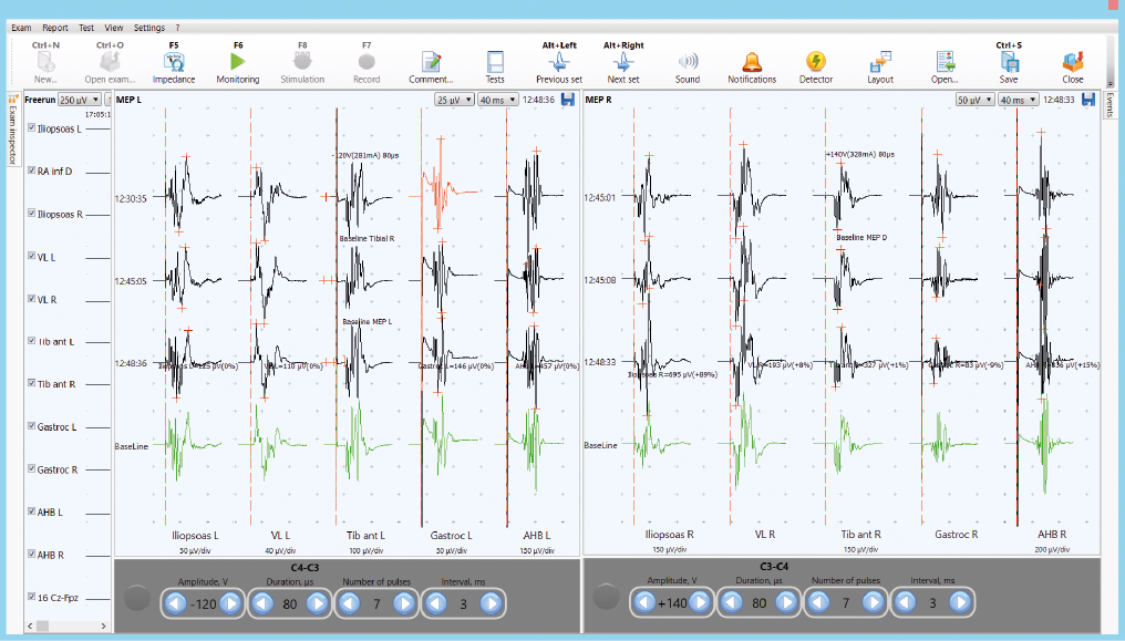
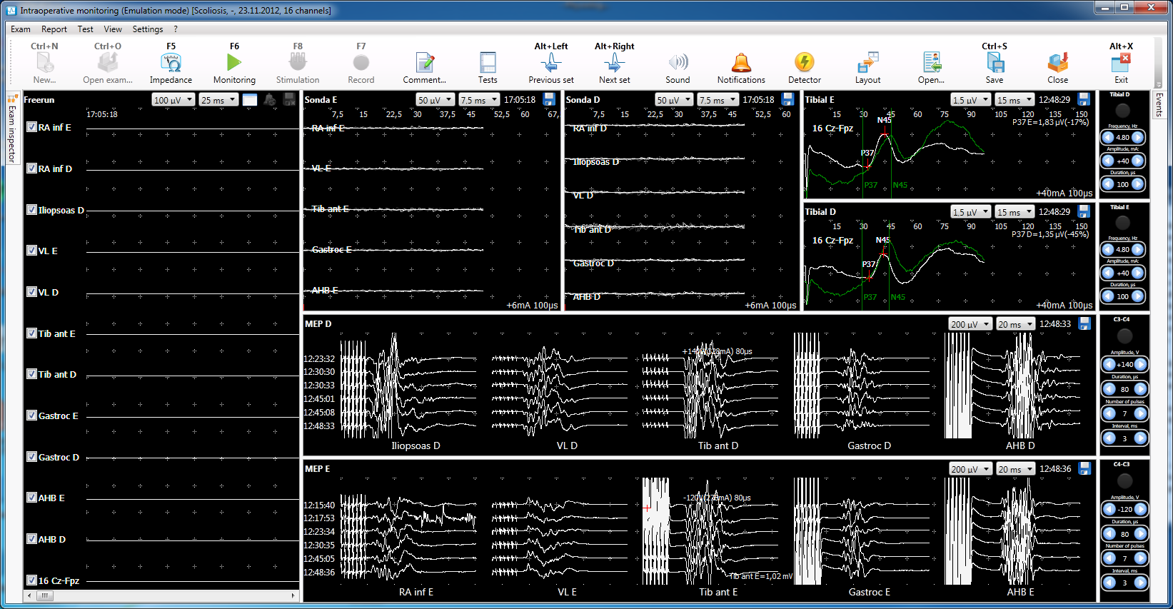
The traces of different modalities can be displayed simultaneously (SSEP, MEP, free-run EMG, etc.). Additionally, it is possible to view only the last obtained traces in “cascade” mode with astronomical time or surgery start time tag. The current traces can be compared with baseline ones.

Data windows image

Video Recording During Surgery

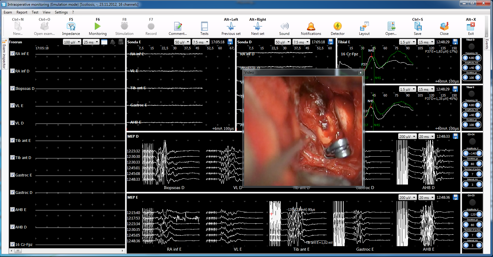
Video from up to three video cameras can be displayed on the screen. The video is recorded and synchronized with other data.

Image of Video Recording during surgery

Test Window Layouts

The possibility to create various layouts of test windows and quickly switching between them allows accommodating a huge amount of information obtained during multimodal monitoring.

Test Window Layouts

“Trends” Window

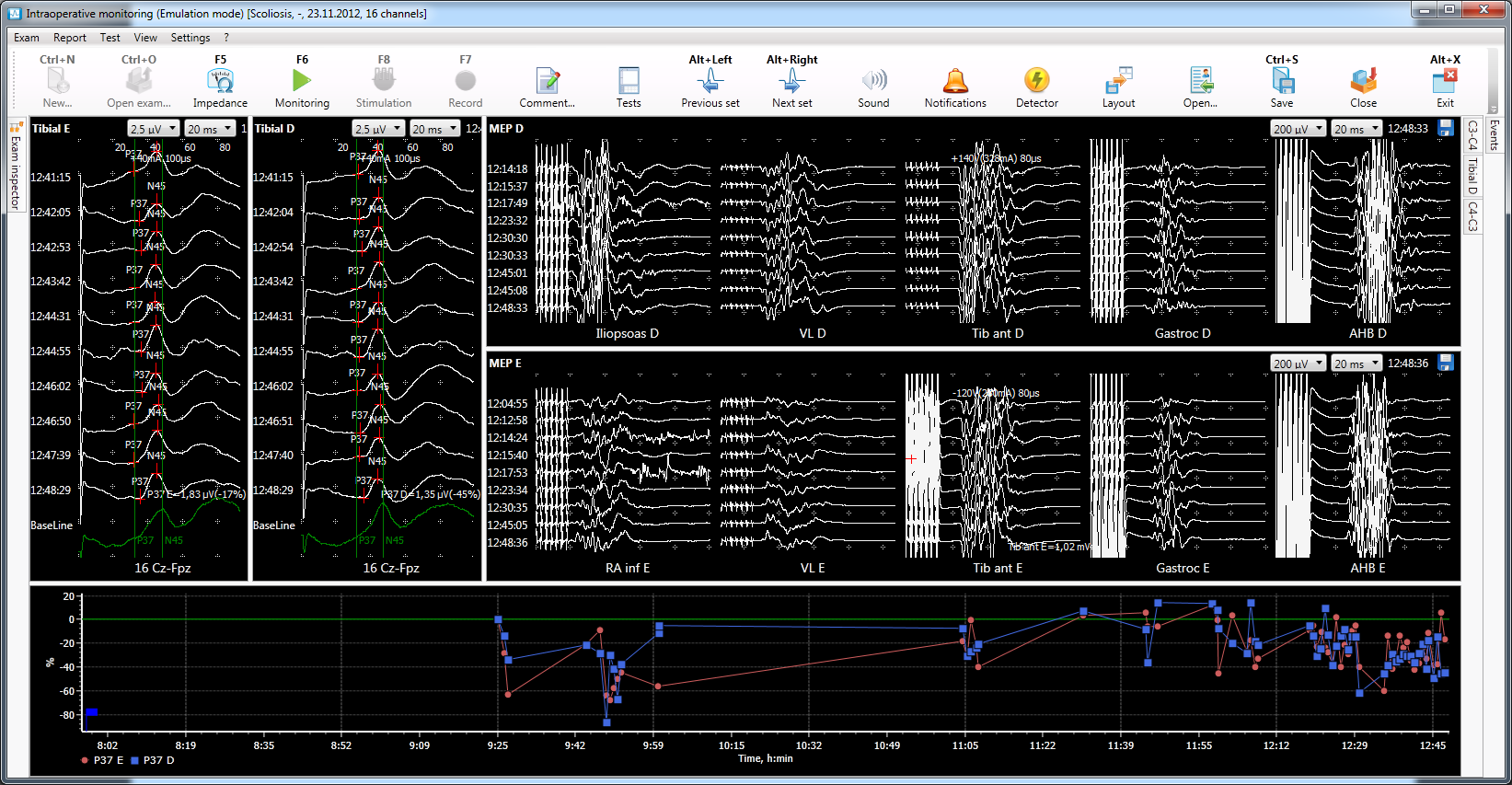
It is possible to view the graphs showing the dynamics during the surgery displayed as trends.

Trends Window Image

Monitoring Report

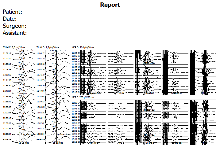
The software allows for automatically generating a monitoring report in .rtf or Word format. The report includes traces and text comments. It can be edited using the built-in editor or Microsoft Word and printed on an office PC printer.

Report Monitoring Image

MEGA-IOM Versions

The system can be supplied in one of three configurations depending on the needs of the neurophysiologists and surgeons. The configurations differ in the number of channels and application at different types of surgeries. The system can be supplied with desktop or laptop computers.

MEGA-IOM 32/B 32/S 16/S
IOM channel 32 32 16
Dedicated channel for routine EMG, NCS, and EP 2 2 2
Electrical stimulator channel 12 4 4
Low current stimulator channel 2 1 1
Transcranial electrical stimulator channel 4 - -
Auditory stimulator channel 2 2 2
ES detector channel 2 2 2
Pattern-stimulator channel 1 1 1
MEGA-IOM by Soterix Medical is United States FDA 510(k) cleared.