Transcranial Focused Ultrasound Stimulation
Low-Intensity Focused Ultrasound Pulsation

Transcranial Focused Ultrasound Stimulation: Low-Intensity Focused Ultrasound Pulsation

Fully integrated solution for rigorous and safe experimentation. Validated compatibility with MRI and fMRI. Only proven placebo solution in industry. EEG synchronization. Unique calibration process for reproducibility. Developed by BrainSonix, the pioneers of tFUS.


About Transcranial Focused Ultrasound

Ultrasound has been used extensively in medicine and industry for more than 70 years (Fry 1958; Rezayat 2016; Darrow 2019) by delivering mechanical energy (sound waves) to tissue. Ultrasound as a technique for neuromodulation is essentially characterized into two main categories segregated by the intensity (spatial peak temporal average intensity [SPTA]) of the acoustic wave. High-intensity focused ultrasound (HIFU) with intensity typically >1 W/cm2 produces reliable and permanent brain lesions through thermal ablation, whereas low-intensity focused ultrasound (LIFU) does not produce lesions but can excite or suppress neural activity with very rare occurrence of adverse effects (Pasquinelli 2019). Although HIFU secured the United States Food and Drug Administration (FDA)’s approval for essential tremor in 2016 and approval for other conditions is apparently on the horizon, systematic efforts aimed at determining clinical utility for LIFU have only recently begun (Truong 2021). Transcranial focused ultrasound (tFUS) is a LIFU technique that is characterized by long trains (several hundred milliseconds) and has been shown to both potentiate and suppress neural activity (Bystritsky 2011; Bystritsky 2015) as well as alter behavior in mammalian brains (Fomenko 2018). The low energy of tFUS falls below the threshold for inducing any tissue damage and is often used with intensities that are near or below those used in diagnostic ultrasound.

System Components

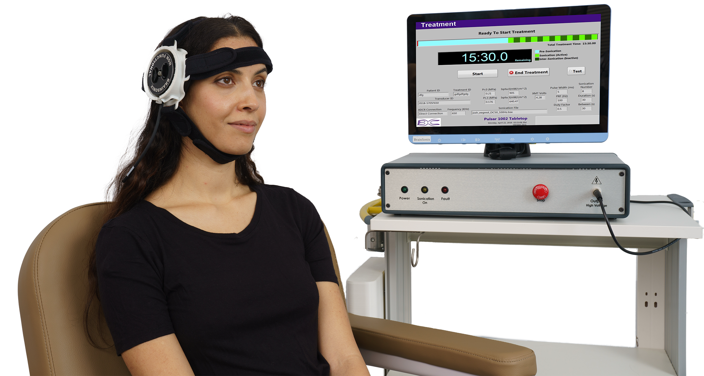
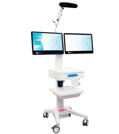
The BrainSonix system comprises the ultrasound console (stimulator), transducer with ground-breaking “clipping” mechanism, transducer holder, and coupling pads. The system is fully integrated (i.e. enclosure and monitor) allowing portability and tabletop operation. The stimulator includes an all-in-one computer with an ultrasonic drive generator. The ultrasonic drive voltage is provided through a BNC port on the front panel. Upon entering the desired exposure parameters (pressure, average intensity, pulse length, pulse repetition frequency, sonication duration, intersonication time, and number of sonications), the system automatically calculates the voltage required to meet those settings (Schafer 2021).

Three different circular, single-element, spherically focused transducers are available with an aperture of 61 mm. Nominal focal lengths of 55, 65, and 80 mm are provided for targeting different regions of the brain. The transducers include a slightly convex front layer for optimal impedance matching.

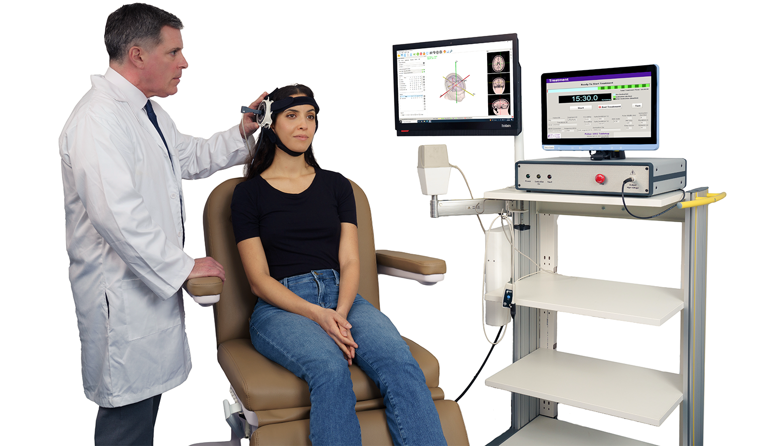
The transducer holder attaches the transducer firmly to the head above the temporal window. It allows both angular and linear displacement to accurately target intended structures.

MR synchronization is provided via a fiber optic input line and optically isolated digital signal line (TTL). EEG synchronization is also provided either via TTL or optical signal.

State-of-the-art calibration, reproducible operation, and uncompromising safety

The main goal of the calibration process is to ensure that the intended ultrasound exposure is provided on a repeatable and consistent basis. The BrainSonix system includes a state-of-the-art calibration technique to ensure that the experimenter can focus on the clinical study and not on replicability (Schafer 2021). Specifically, the process is conducted for every stimulator, in combination with any specific transducers, cables, RF filters that are supplied with the system. The calibration settings are stored in a file and made available for post-hoc stimulation verification.

When setting up for a stimulation session, calibration data is first retrieved. The user is first asked to confirm the transducer serial number and cable configuration. The system then obtains the acoustic output characteristics from the calibration file data. Next, the user is asked to enter desired stimulation parameters. Based on acoustic output limits established for the study protocol, the system automatically determines whether the entered treatment parameters exceed limits. The system will not allow proceeding to the stimulation initiation step, if parameters are not adjusted to be in compliance with output limits.

Certified, validated technology with an extensive track record with regulatory agencies

The BrainSonix device has been rigorously validated in several clinical trials. The system has secured multiple Investigational Device Exemption (IDE) and Non-significant Risk (NSR) letters from the United States FDA. The system is IEC electrical safety and EMC certified. Contact Soterix Medical team for any support related to trial approvals (IRB / IDE in the United States and ITA in Canada).

Validated compatibility with fMRI

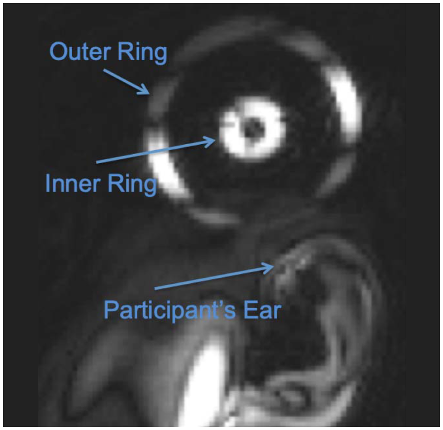
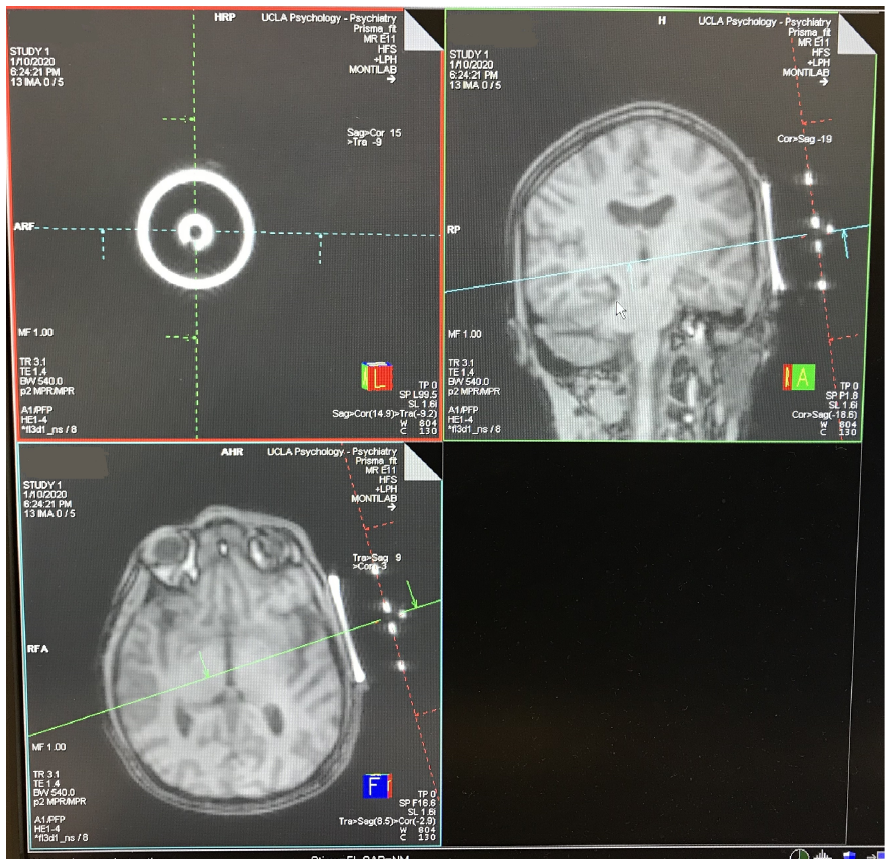
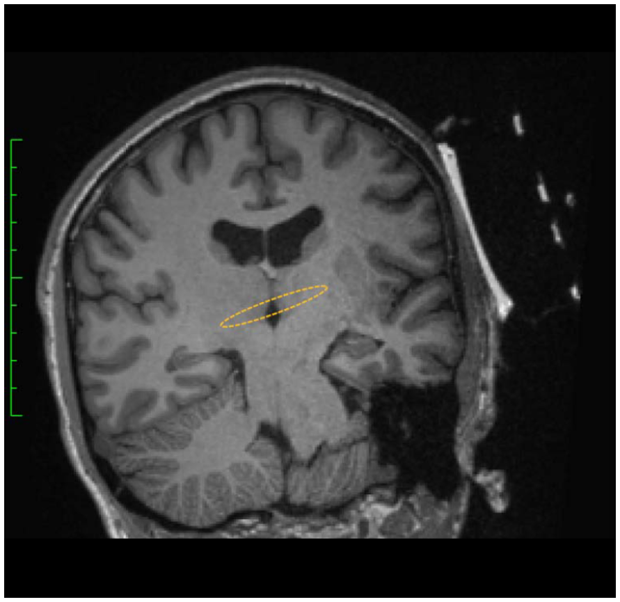
MR compatibility is typically complex for any tFUS system, as transducers contain electrically conductive materials. BrainSonix transducers have been specially designed with internal MRI-lucent fiducial materials and subsequently validated for safe operation in a 3T MRI environment. Specifically, no interaction at 128 MHz with the scanner RF transmit and receive coils was demonstrated (Korb 2014). The transducer is interfaced with a MR conditional cable and connected to the stimulator console through a wall filter. The provided transducer holder is MR safe that allows for simple and robust positioning and angulation. MR synchronization is provided via a fiber optic input line and optically isolated digital signal line (TTL) allowing simultaneous MRI acquisition. Image on the left below shows MRI image of transducer fiducial markers. Image in the middle below shows the alignment process. Image on the right below shows the approximate position (yellow oval) of the focal zone (Schafer 2021).

Only proven sham / placebo solution

Transducers produce audible sounds and detectable vibrations making blinding a major confound in tFUS trials. In 2023, BrainSonix team engineered a special matched set of acoustic coupling pads that either pass or block the ultrasound wave (Patent Pending). These pads appear visually identical to both subject and operator. The true property of each pad can be disclosed to the independent study coordinator or the statistician based on the clinical study plan. Contact Soterix Medical for your trial-specific placebo control plan.

Neuronavigation Integration

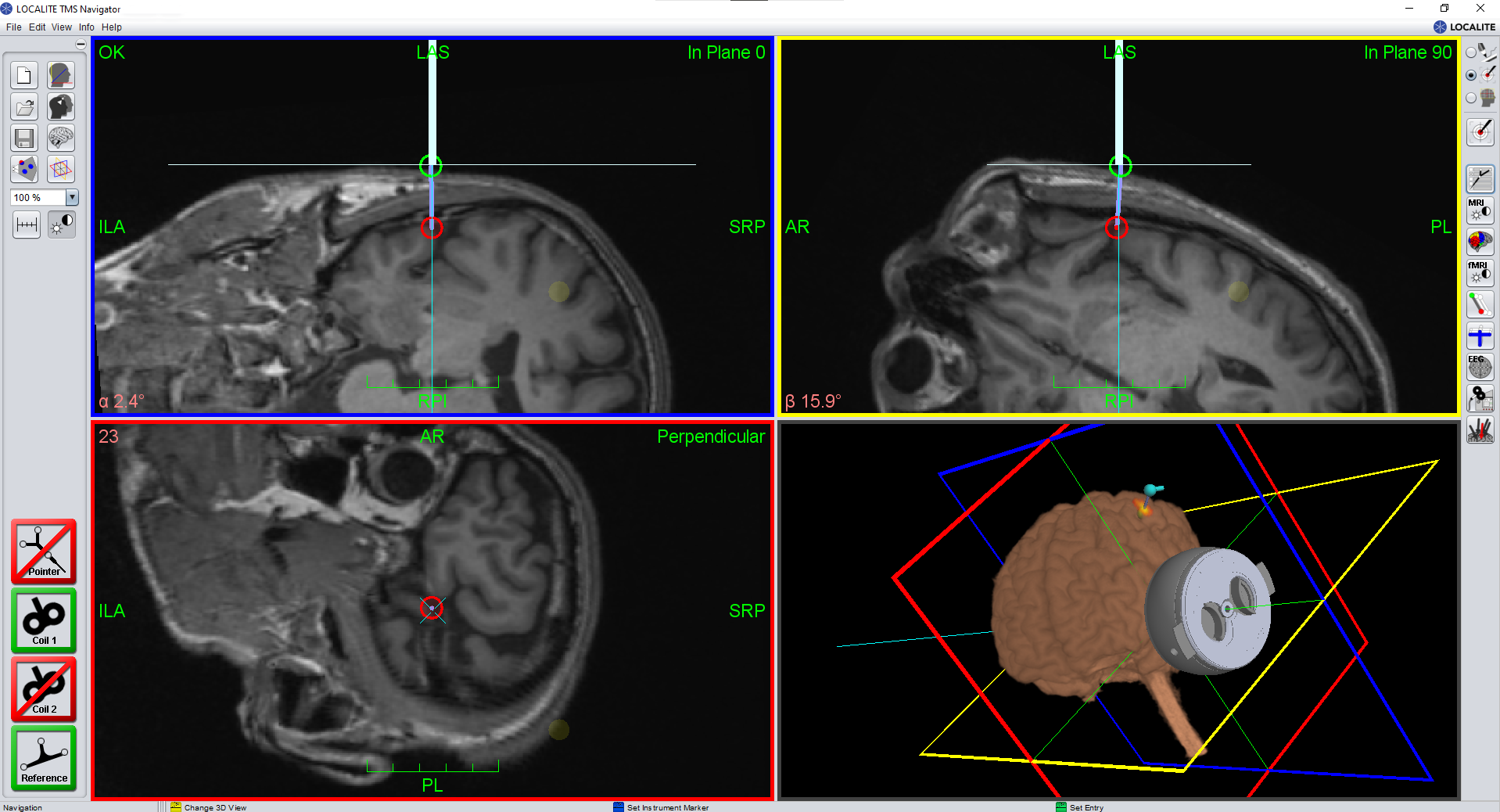
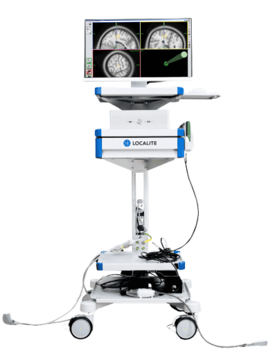
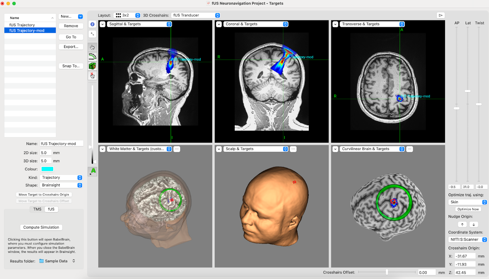
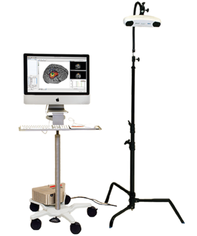
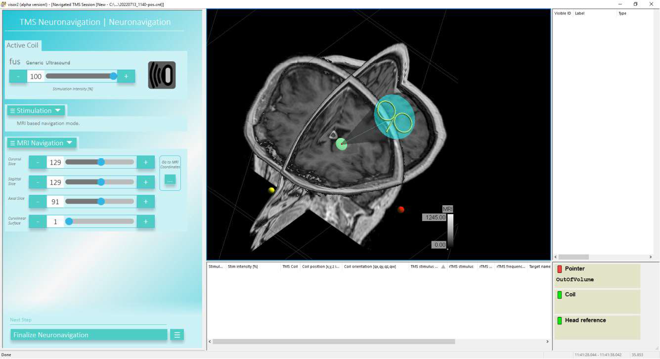
Navigation tracking is often critical in tFUS to confirm whether the focal zone is actually located within the intended brain target. The BrainSonix device has been validated to work with either magnetic or optical based tracking systems. Solutions currently exist for integrating with BrainSight, Visor 2, and Neural Navigator neuronavigation solutions.

If you are interested in pairing with your neuronavigation solution, contact Soterix Medical for additional information.

References

  1. Production of reversible changes in the central nervous system by ultrasound. Science. 1958;127:83–84. Fry FJ, Ades HW, Fry WJ.
  2. A review of low-intensity focused ultrasound pulsation. Brain Stimul. 2011 Jul;4(3):125-36. Bystritsky A, Korb AS, Douglas PK, Cohen MS, Melega WP, Mulgaonkar AP, DeSalles A, Min BK, Yoo SS.
  3. Low-intensity focused ultrasound pulsation device used during magnetic resonance imaging: evaluation of magnetic resonance imaging-related heating at 3 Tesla/128 MHz. Neuromodulation. 2014 Apr;17(3):236-41; discussion 241. Korb AS, Shellock FG, Cohen MS, Bystritsky A.
  4. A Review of Low-Intensity Transcranial Focused Ultrasound for Clinical Applications. Curr Behav Neurosci Rep 2, 60–66 (2015). Bystritsky, A., Korb, A.S.
  5. A Review on Brain Stimulation Using Low Intensity Focused Ultrasound. Basic Clin Neurosci. 2016 Jul;7(3):187-94. Rezayat E, Toostani IG.
  6. Focused Ultrasound for Neuromodulation. Neurotherapeutics. 2019 Jan;16(1):88-99. Darrow DP.
  7. Safety of transcranial focused ultrasound stimulation: A systematic review of the state of knowledge from both human and animal studies. Brain Stimul. 2019 Nov-Dec;12(6):1367-1380. Pasquinelli C, Hanson LG, Siebner HR, Lee HJ, Thielscher A.
  8. Design, Development, and Operation of a Low-Intensity Focused Ultrasound Pulsation (LIFUP) System for Clinical Use. IEEE Transactions on Ultrasonics, Ferroelectrics, and Frequency Control. 2021 Jan;68(1):54-64. Rezayat E, Toostani IG.
  9. Comparison of Transcranial Focused Ultrasound and Transcranial Pulse Stimulation for Neuromodulation: A Computational Study. Neuromodulation. 2022 Jun;25(4):606-613. Truong DQ, Thomas C, Hampstead BM, Datta A.
Caution! Investigational Device. Federal (or United States) law limits device to investigational use.