Worldwide
LoginSoterix Medical is tackling depression with a drug-free approach. Navigated rTMS ensures that treatment pulses are precisely and reliably delivered to the intended target at all times.
Neural Navigator Hardware
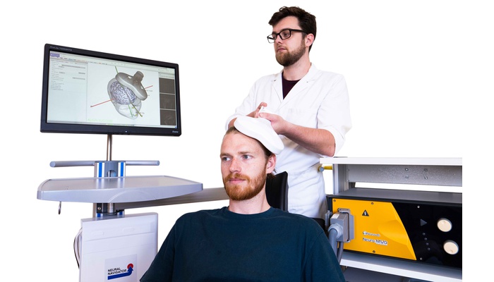
The core principle and effectiveness of the Neural Navigator begins with the hardware. True electromagnetically guided neuronavigation, not hindered by line-of-sight (LoS) issues, is made possible by the magnetic field transmitter. Just as important, are four sensors that each serve a critical purpose. The first sensor is inserted into a coil clamp, used to accurately reflect the position of the coil in respect to the transmitter. The second sensor is in the pointer, used to precisely capture facial landmarks on the patient’s head. The patient wears a headband containing the final two sensors used for head movement tracking.
An ergonomically designed trolley compliments the Neural Navigator. The trolley offers a height-adjustable monitor, allowing pertinent information to be within eye-level of the user. An extended and rotatable arm allows the magnetic field transmitter to have flexibility in position relative to the patient, important for tracking the sensors and target brain regions. In order to ensure that the whole setup is compact during use, the Neural Navigator unit along with the accessories are discreetly attached to the trolley.
Neural Navigator Software
Transcranial Magnetic Stimulation (TMS) requires accurate placement of the TMS coil over the targeted brain area. Conventional placement methods in TMS are based merely on external landmarks of the head (5 cm rule, 10-20 EEG). This leads to inaccuracies in targeting of this brain area and therefore to suboptimal results in TMS research and therapies, and high variability of treatment outcome for different patients.
To overcome this problem, MRI-guided neuronavigation is developed which provides added value to TMS stimulation for more precise, real-time neuronavigation of TMS pulses. Neuronavigation aligns the MRI scan acquired at an earlier point in time with the head of the patient, by measuring several fiducial points on the head with a tracker. At the same time, the TMS coil and the head of the patient is tracked. This allows the neuronavigation software to accurately guide a TMS pulse to the intended neuroanatomical structure with millimeter precision.
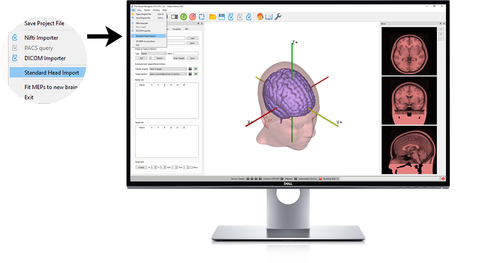
Standard Head Import
The Neural Navigator supports the option to import a MNI-152 standard head model that can be used as a substitute for a patient MRI, allowing a clinic to accommodate any situation where one is not available. The standard head can be warped to fit the patient’s own anatomy. Alternatively, a T1 weighted structural MRI scan can be manually loaded using the DICOM or Nifti import feature within the Neural Navigator.
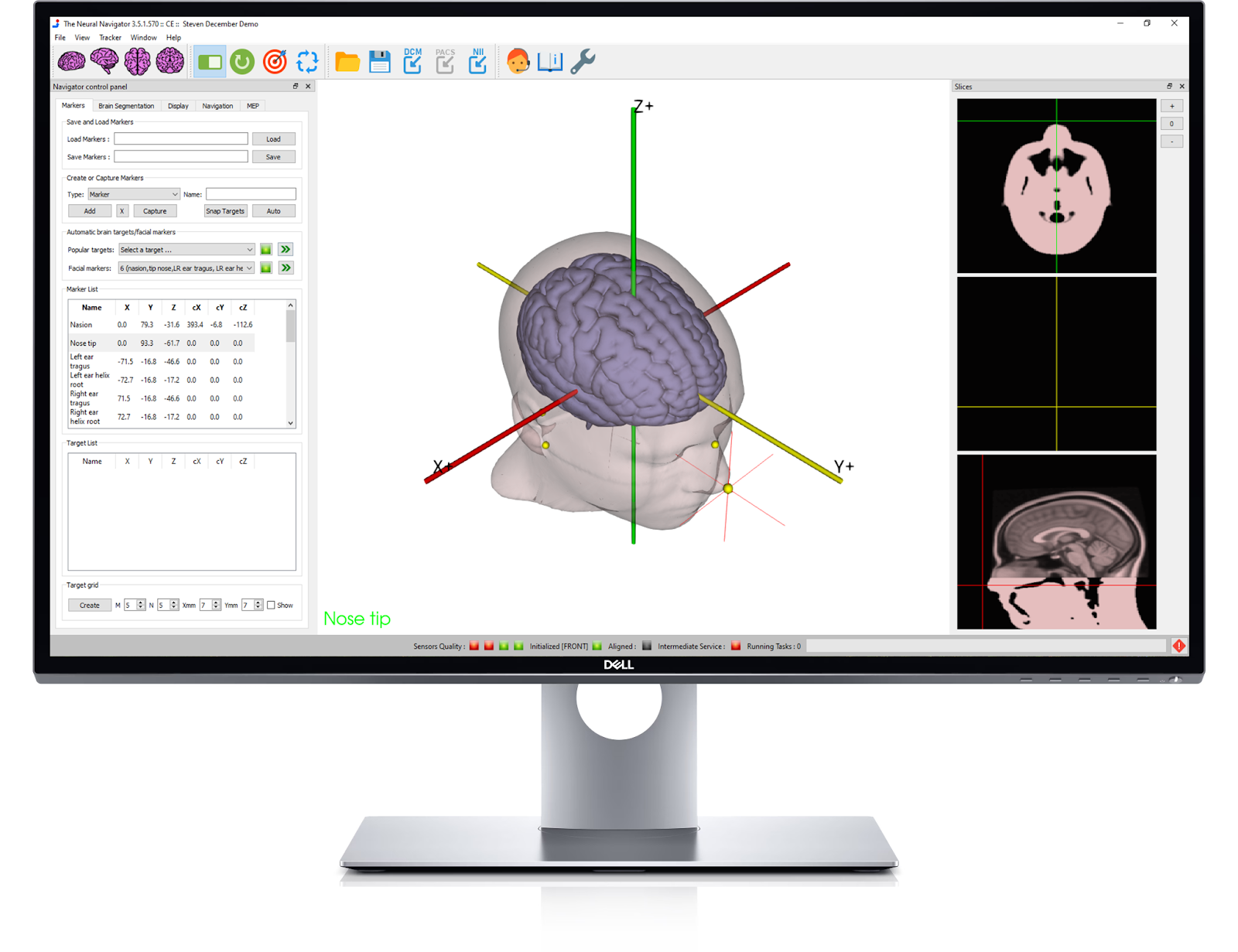
Capturing Facial Landmarks
The Neural Navigator will warp the head model to the patient’s anatomy through the process of collecting anatomical facial landmarks on the patient. Selecting a set of facial landmarks from an extensive list will prompt you to subsequently capture them using the pointer. Text on the screen and auditory cues serve as a guide throughout the anatomical landmark collection process.
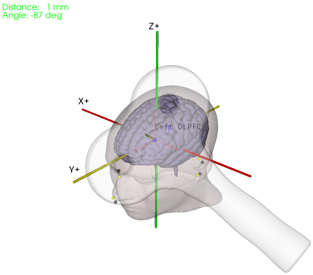
Navigating Coil to Desired Target
Select a target brain region from an extensive list of popular targets. The Neural Navigator software will then display the targeted brain region on the warped head model. Users may use distance, depth, and angle measurements as tools to accurately aim the coil at the targeted brain region. Neural Navigator provides seamless integration and support for a wide range of coils.
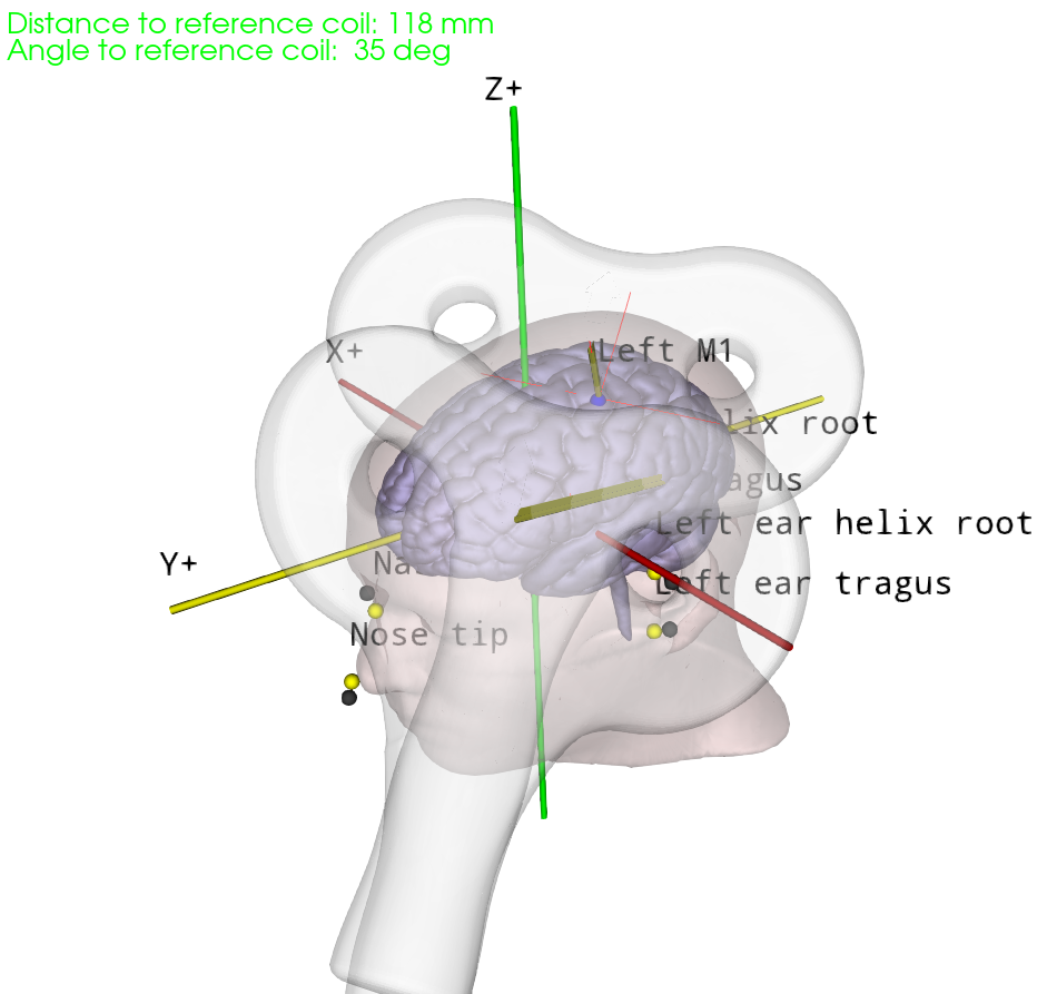
Coil Reproducibility
The Neural Navigator supports accurate reproduction of TMS coil positions using an MNI template. When a target brain region is stimulated, the TMS coil position and orientation are stored. During a follow-up session, this data can be loaded, and the TMS coil position and orientation can easily be reproduced for optimized brain stimulation. Visualization of the reference coil position and distance and angle indicators help guide the TMS coil to the reference position.