Soterix Medical Neuronavigation

Neuronavigation to precisely and accurately position your TMS coil or electrodes for HD-tES

The only navigation system that is not affected by line-of-sight occlusion issues without compromising precision and accuracy

FDA cleared for rTMS navigation, increasing reliability and reducing set-up time

Fully integrated EMG recordings for unmatched precision in mapping functional brain motor areas

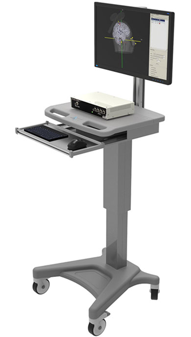
TMS Neuronavigation (Essentials Version)

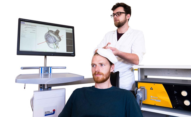
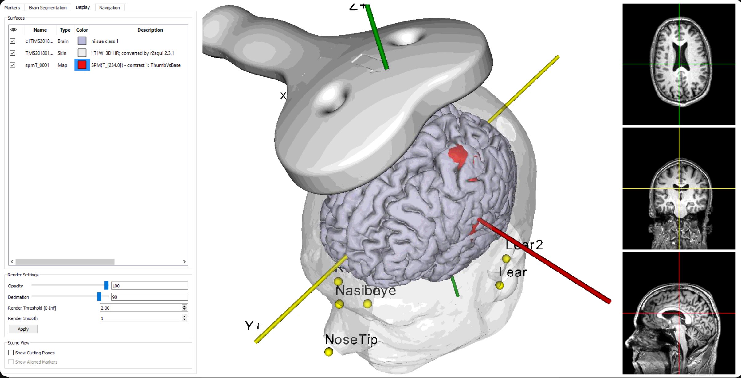
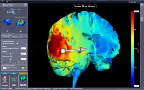
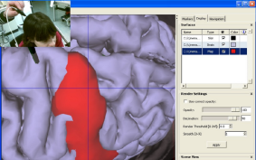
TMS induces focused electrical current in desired brain regions via brief, time-varying magnetic fields produced by a coil which is typically hand-held. The precise and reliable positioning of this coil is not a trivial task. A millimeter displacement from a specified target location may potentially compromise your results. Further, no two brains are alike; therefore coil placement simply based on head shape alone (‘5cm rule’) or the International 10-20 EEG is often ineffective. The Neural Navigator navigation system provides unmatched precision in positioning the TMS coil over a specified target based on an individual’s MRI. Desired brain targets can be identified by manually selecting them or by combining with fMRI determined regions of activity in real time.

TMS Neuronavigation

TMS+EMG Neuronavigation (Navigated MEP version)

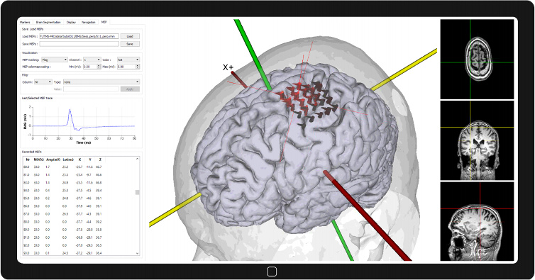
The Neural Navigator Navigated MEP (NavMEP) integrates EMG recordings with the high precision navigation system of the Neural Navigator. The Neural Navigator NavMEP is designed to map the functional motor area onto the brain surface using MRI-guided neuronavigation, monophasic Transcranial Magnetic Stimulation (TMS) and simultaneous electromyography (EMG) recordings. The functional motor maps can be used for monitoring of neural changes during stroke rehabilitation. The NavMEP records the stimulated brain region, the stimulation intensity and the features of the motor-evoked potential. It integrates this information and presents it as an intuitive functional motor map onto the brain surface. The NavMEP offers high precision neuronavigation integrated with focal monophasic TMS and high quality EMG recordings. The software contains a built-in segmentation algorithm in order to obtain the brain surface from raw MRI images. The neuronavigation device allows uninterrupted tracking of both the TMS coil and the head.

TMS + EMG Neuronavigation (Navigated MEP)

HD-tES Neuronavigation

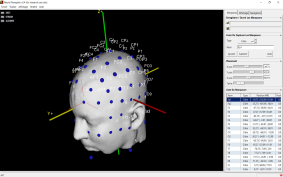
Soterix Medical High-Definition transcranial Electrical Stimulation (HD-tES) is a non-invasive technique where desired brain regions are targeted using arrays of scalp electrodes. Soterix Medical neurotargeting software, HD-Targets, leverages mathematical optimization to automatically determine the best HD electrode configuration for any target. For patient-specific optimization, localizing the HD sites onto the patient’s scalp based on patient’s MRI ensures precise electrode placement. This as a result ensures precise and consistent HD-tES application.

Perform HD-tES Neuronavigation in three easy steps:

1) Use HD-Targets to determine optimal electrode placement for your brain target.

2) Load stimulation electrode voxel locations into Neural navigator software.

3) Navigate to stimulation electrode location using individual's MRI and mark location on scalp.

Why is immunity to line-of-sight occlusion is important?

Most navigation systems use either optical (infrared) or electromagnetic tracking technology. Both systems perform the same function- however, the technology used to provide information to the operator is substantially different. The optical system uses infrared sensors in combination with light emitting structures that are fixed to the subject’s head via a headband and fixed to a hand-held probe. Both the headband and the instrument must be “seen” by the system’s camera in order to allow navigation. Optical systems are typically more expensive. The electromagnetic systems like the Neural Navigator on the other hand use electromagnetic fields that use reference points on a device attached to the patient’s head (held via headband). Unlike the optical systems, electromagnetic systems do not have to be “seen” by a camera and therefore do not suffer from line-of-sight (LOS) occlusion issues. In fact, electromagnetic tracking dominates the surgical navigation field due to no LOS occlusion issues. Furthermore, the Neural Navigator system uses DC pulsed tracking enabling it to match the precision and accuracy of optical systems.

The net result therefore is a fundamental advance in Neuronavigation – a system that matches the precision and accuracy of optical systems while not being affected by LOS occlusion that is more affordable.

Electromagnetic versus Optical Tracking

During neuronavigation, the 3D positions and angles of the TMS coil, a digitizing stylus and the head of the patient typically must be known at all times. Position tracking of coil, head and stylus can be performed either by using optical position tracking with large cameras on a stand or by electromagnetic (EM) position tracking based on a DC pulsed magnetic field emitted by a small box. In neurosurgery, where the principle of frameless stereotactic neuronavigation was invented decades ago, both optical and EM tracking techniques have been around for a long time and have proven their worth. They are found to be equally accurate in larger studies (Koivukangas 2013 & Ricci 2008). Especially EM tracking offers several advantages and is currently popular in neurosurgical navigation applications requiring high precision and reliability. The main advantage of EM tracking remains immunity to line-of-sight occlusion, or avoiding the inability to track and navigate when the camera view is blocked, for example by the arms of a person using the equipment. However, another important advantage is the compact nature of EM tracking, no big camera stand is needed, and sensors are much smaller as well. The DC pulsed EM navigation that neurosurgeons use often and is also used in the Neural Navigator is very robust as it does not induce so called ‘Eddy currents’ in metals that otherwise would cause distortions. The material used inside a TMS coil also does not pose any problem for EM position tracking based on DC pulses.
The Neural Navigator was originally developed and rigorously tested at Utrecht University Medical Center in the Netherlands currently has numerous users around the globe – some of which have been successfully using the system since 2011. Of course a magnetic pulse generated by a TMS coil very briefly interferes with EM position tracking nearby the TMS coil, for about 1 millisecond. This brief distortion is detected in real-time and removed by the Neural Navigator proprietary and fully validated algorithms. This allows seamless position tracking of coil and head even during high frequency rTMS and even theta burst TMS protocols.

Navigated TMS is significantly better than the 5 cm rule or 10-20 method

No two brains are alike, making precision placement of the coils used in TMS Therapy tricky. Precise and reliable positioning can make the difference between a successful result and an unsatisfactory one. Conventional coil placement is done using anatomical landmarks like the bridge of the nose, the projecting part of the back of the skull, etc. to align the coil over an intended brain target. This is called the “non-navigated approach”, which does not require the patient’s MRI.

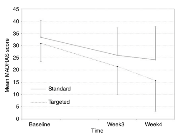
The “navigated approach”, on the other hand, uses the patient’s MRI, thereby taking into account the individual’s anatomy. From the MRI, one marks out desired brain targets using individual brain landmarks (gyri and sulci). Once marked, the coil can be positioned to within a mm precision over the target. Any subject or operator movement can be immediately compensated for- as the brain target is always visible. This approach is similar to the one taken in image-guided stereotaxy when performing brain surgery. Studies have shown unequivocally greater benefit (up to twice as much) when using a navigated approach over the non-navigated one for TMS treatment.

Navigated TMS

Figure 2. Change in Montgomery-Asberg Depression Rating Scale (MADRS) scores (least square means) over time.

Figure on left from "A randomized trial of rTMS targeted with MRI based neuro-navigation in treatment-resistant depression” Fitzgerald PB, Hoy K, McQueen S, et al. Neuropsychopharmacology 2009.

‘Standard‘ indicates Non-Navigated approach

‘Targeted‘ indicates Navigated approach

Features

  • Precision of 4mm or better.
  • Spatial accuracy better than 1 mm.
  • Universal TMS compatibility with coil sockets available for Magstim, Magventure, and Neurosoft TMS coils. Customization available for TMS coils from other manufacturers.
  • Import of standard MRI data types (Nifti, Analyze, and DICOM).
  • 3D MR constructed scalp, brain and activation (fMRI) data.
  • Intuitive software workflow.
  • Complete setup fits in a small suitcase making it ideal for bed-side investigation and easy transportation.

Resources

Upon request, Soterix Medical support team can provide assistance and advice in MR image processing and MRI data quality inspection.

Neural Navigator by Brain Science Tools BV is exclusively distributed in the US and Canada by Soterix Medical. Neural Navigator has extensive regulatory agency approvals worldwide: Neural Navigator is United States FDA 510(k) cleared. Neural Navigator is Canada (Health Canada) Licensed.

The only neuronavigation system that is fully compatible with high definition transcranial electrical stimulation


Learn more about Soterix Medical HD-tES